X-rays are safe and necessary tools to aid the dentist in diagnosis and successful treatment.
In this column, we will discuss the importance and benefits of dental x-rays. X-rays utilize modern technology to visualize inside the body for evidence of disease and to gain further information that we cannot see with the naked eye.
X-rays are noninvasive and painless. They are used to assist is diagnosing infections, disease, bone loss, bone gain and monitor therapy to predict outcomes.
In general medicine, x-rays support medical doctors and technicians in the use of catheters, stents or other devices inside the body, treat tumors or remove blood clots or other blockages. In essence, we cannot function without accurate x-rays.
Also, x-rays can help to determine the proper course of treatment and reveal underlying disease or infection. For example, patients often come to the dentist complaining of a tooth abscess and severe pain. Looking in the mouth the abscess appears to be a gum abscess however upon closer examination the x-ray shows the actual source of the problem- nerve damage and infection inside the tooth. This is important because the treatment varies depending on the cause of the infection.
In dentistry, we use several types of x-rays. They include Periapical X-rays, Bite wings, Panoramic imaging or 3-D Cone Beam (CT Scan). The Peri-apical and Bitewing x-rays are staples in the dental industry. Today, the Panoramic and 3-D CT Scan have evolved as game-changers because the dentist is now capable of seeing so much more. In essence, the guess work is over. We can now better predict treatment out-comes.
The modern x-rays are digital. This means they are high quality definition. They can be transported via the internet and there is no need for chemicals in processing. Digital x-rays utilize less radiation, therefore are safer. Digital x-rays reduce radiation exposure for both the patient and the dentist up to 90%.
Digital x-rays are less expensive to process yet are more accurate than traditional ones. The digital x-ray image instantly projects onto the dentist’s computer. It can also be sent via the internet to any computer or cell phone.
A 3D Cone Beam dental scan shows multiple images in different planes. It is better for identification of disease or pathology. The 3D scan allows the specialist to see the correct volume and thickness of bone or soft tissue which is critical for assessing bone volume for a dental implant. Proper examination of x-rays and CT scan come with considerable training and experience which enables the dentist to give an accurate diagnosis and effective treatment plan.
Recently a patient came to my office with discomfort because they suffered from wearing a poorly fitted lower denture. They were discouraged because they were advised their lower jaw could not take implants. The patient felt helpless and dejected.
As a part of the comprehensive examination, a CT scan was taken. After assessing the precise location of the important structures on the lower jaw we were able to assist this patient by placing four dental implants and ultimately restoring their smile. Today the patient is happy and smiles often with their new teeth.
X-rays are a reliable asset to the dentist thus providing a great service to patients. They are a necessary tool in diagnosing and treating simple and complex conditions. Remember, a dentist cannot fix what they cannot properly see.
Dr. Kendal V. O. Major is Founder and CEO of Center for Specialized Dentistry which is a comprehensive family dental practice operating in Nassau and Freeport. He is the first Bahamian Specialist in Gum Diseases and Dental Implants since 1989. He is a Certified Fast Braces Provider. His practice is located at 89 Collins Avenue, Nassau at (242)325-5165 or [email protected].

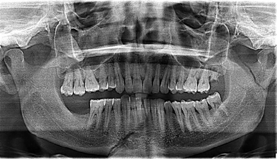
Panoramic x-ray- See dropping (extrusion) of molars due to early loss of lower molars

Cone Beam CT Scan shots. A 3-dimensional imaging that allows the dentist/radiologist to see vital structures and measure volume and thickness in axial, frontal and sagittal planes

Computerized Digital Image of a Peri-apical x-ray. Note the very dark areas representing infection or blood vessels. The white are metal fillings and post/core root canal.


